This is a sample of the report - note outline of bones is demonstrated in radiographic positions section.
Good radiographic positioning.
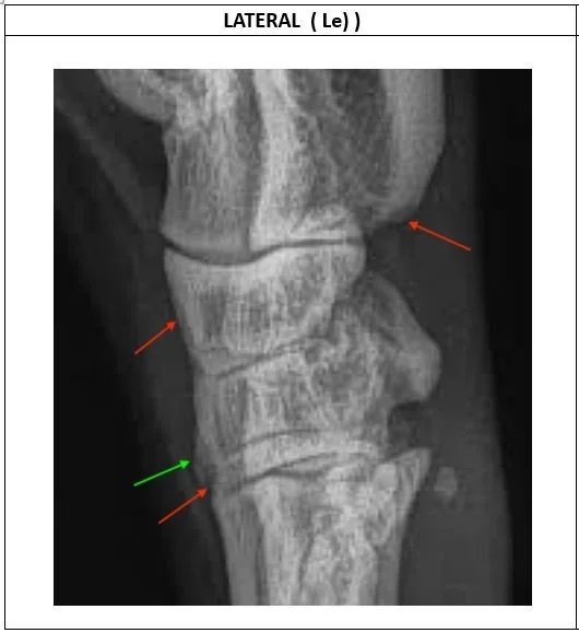
Plantar lesion – red arrow points to cortical lysis of calcaneus. There is hint of soft tissue calcification just distal to this arrow. This may indicate that the plantar ligaments have been damaged and are now over-contracted.
Dorsal (forward facing) lesions include
lysis in cortex of CTB (red arrow)
excess callus base of T3 (green arrow)
lysis at interface of T3 and MT3 (red arrow).
These changes in the dorsum may indicate that there is overload due to weight and increased torsional strain. This may occur with laxity of the dorsal soft tissues.
Good radiographic positioning.
Mild cortical lysis in distal talus (red arrow) which may be due to over-stretching of soft tissues and is common but rarely any problem.
Green arrow points to increased whiteness (sclerosis) in top of metatarsal III which is associated with vertical weight and torsional stress.
Good radiographic positioning.
Green arrow points to presence of distinct corner of the CTB which indicates good calcification and no bone loss.
The radiographic positioning could be improved by being only 30 degrees from the standard PD position. This view is more like 60 degrees.
There is a vertical fracture line in T4 (marked with red arrows).
Report Summary:
Acute pain on front (dorsum) of left hock is likely due to the vertical crack in the T4 bone.
The fracture is not complete.
Note: there are pre-existing lesions due to overload within the tarsal structure in the plantar and dorsal zones.
Risk Assessment: Low/ Medium
Recommendatons:
Vertical fracture in T4 may heal with 6 weeks rest but advise repeat radiography at 3-4 weeks to confirm progress.
The fracture line may require compression with surgical screw insertion (Katakasi, AGWSDV conference 2023). Advise to discuss further with Dr Katakasi at Adelaide Plains Veterinary Surgery.
Rest will also assist calcification in the dorsal areas of the CTB, T3 and MT3.
To reduce future overload in the soft tissues:
Strapping may counter the laxity of the dorsal tissues.
Injections into the plantar ligaments may reduce load on the calcaneus.
DISCLAIMER:
All efforts are made to provide an accurate report, however, at times, not all lesions are observable in radiographic images. The above recommendations are made in good faith and with the intention of providing education to minimise the risk of hock fracture.
The recommendations made by the veterinarian, are in no way exhaustive and only part of the requirement for a treatment regimen. The judgement of the owner / trainer of the greyhound also influences the decisions that are made in the follow up to this report.
The veterinarian cannot take responsibility for any lesions that are not evident in the images provided, nor for any treatment administered at the discretion of the owner /trainer.
All recommendations made in this report should be discussed with your local veterinarian.


